Abstract
Ovarian cancer ranks among the most frequently diagnosed gynecological malignancies during pregnancy.1 Prevalence in general population is 1 in 15000 to 1 in 32000 pregnancies.2 An estimated 2.8 to 11 per 100,000 pregnancies are complicated by ovarian malignancies, with approximately 5% of these tumors being cancerous. Despite the low incidence of gestational ovarian malignancy, it affects maternal and perinatal management.3Tumors persisting in 2nd trimester, complex or greater than 6cm should be managed surgically.
Keywords
Juvenile Granulosa Malignancy
Ovarian cancer ranks among the most frequently diagnosed gynecological malignancies during pregnancy.1 Prevalence in general population is 1 in 15000 to 1 in 32000 pregnancies.2 An estimated 2.8 to 11 per 100,000 pregnancies are complicated by ovarian malignancies, with approximately 5% of these tumors being cancerous. Despite the low incidence of gestational ovarian malignancy, it affects maternal and perinatal management.3Tumors persisting in 2nd trimester, complex or greater than 6cm should be managed surgically.4
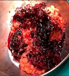
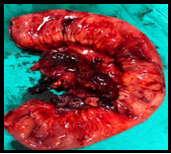
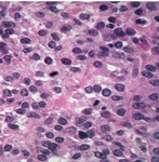
We report a case of A 18year old female, married for 1.5years, Primigravida at 20+4weeks presented in the Emergency department of Obstetrics and Gynaecology Services Hospital Lahore. The presenting problems were abdominal pain, abdominal distension from 8days and constipation from 4days. Physical examination reveal abdomen tense, distended, fluid thrill +ve. On Ultrasound there was 15cm adnexal mass with solid and cystic components and abdominopelvic ascites; possibility of ovarian malignancy. Provisional diagnosis was Malignant Ovarian Tumor. An exploratory laparotomy (Hysterotomy followed by TAH+BSO+Infracolic omentectomy and resection-anastomosis) was performed, confirming Stage IVB Malignant Ovarian Tumor with gross metastatic spread to the liver surface and mesentery (Fig.1). On gross examination, Rt.ovarian tumor 18 * 20cm with torsion. Whole tissue was necrosed, friable, 4-5 litres of hemorrhagic ascitic fluid drained(Fig.2). Metastatic deposits noted in mesenetry, anterior surface of bladder, pouch of douglas, omentum was thickened & fibrosed. Liver was granular with metastatic deposits. Multiple mesenteric and para-aortic lymph nodes largest one 2.5cm noted in the mesentery. Tumor deposits on distal ileal mesentery and enlarged lymph nodes (Fig.3). Resection anastomosis of distal ileum done 8cm from ICJ, taking down about 15cm of ileum alongwith mesentery. Frozen section sent which revealed Neoplastic Ovarian Mass Suspicious for Malignancy. Microscopic examination reveals a neoplasm arranged in sheets intermixed with follicle like structures filled with mucoid material. The cells are small and cuboidal to polygonal in shape, with round hyperchromatic nuclei, noticeable nucleoli, and scant cytoplasm. Areas of infarction are prominent multiple mitotic figures (>20 mitosis/10HPF) ( Fig.4), 4 reactive lymph nodes identified with Immunostains Inhibin and Calretenin positive in tumor cells. Final Diagnosis: Sex cord-stromal tumor; Juvenile Granulosa Cell Tumor, FIGO Stage IVB.
The management of Granulosa Cell Tumor is based on surgery. Multidisciplinary approach is required between gynecological oncological surgeons, obstetrician, radiologists, oncologist, anesthesiologist, neonatologist and pathologist. Conservative management is for early-stage tumors. A special emphasis on psychological and sexual counselling for management and good quality of life. For enhanced surgical approach regarding Granulosa Cell Tumor more studies are required to predict fertility, oncological results and prognosis of patient.

References
- Kaja M, Aneta CP. Approaches to the Diagnosis and Management of Ovarian Cancer in Pregnancy. Cancer Manag Res. 2021;13:2329-2339. DOI ↗ Google Scholar ↗
- Dominik DF, Radzislaw M, et al. Ovarian Cancer and Pregnancy- A Current Problem in Perinatal Medicine: A Comprehensive Review. Cancers. 2020;12:3795. DOI ↗ Google Scholar ↗
- Guidi S, Berghella V, et al. Adult Granulosa Cell Tumor in Pregnancy: A New Case and a Review of the Literature. Healthcare. 2021;9:1445. DOI ↗ Google Scholar ↗
- Aymen FM, Majed G, Amin B, et al. Advanced Granulosa Cell Tumor and Pregnancy: A Case Report, How to Treat and How to Preserve Fertility? Endocrinol Metab Syndr. 2016;5:250. Google Scholar ↗